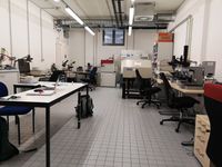
AFM LAB
Our AFM-Lab offers a wide variety of microscopic methods to access the internal morphology of cells, tissues or even whole organisms. Apart from classical light microscopic approaches (brightfield, darkground, phase contrast, polarization, fluorescence and differential interference contrast (DIC)), the lab is also equipped with four atomic force microscopes (AFMs), offering state-of-the-art ultrastructural resolution, high reliability and low maintenance costs.
Additionally, several microtomes (e.g. Ultramicrotome Reichert Ultracut E) enable us to cut virtually any biological material down to 50 nm slices in order to elucidate the samples´ ultrastructure by AFM and light microscopy.
AFM-Lab AK Herrmann (Institute of Pharmaceutical Biology and Phytochemistry)
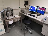
Asylum Research Cypher-S AFM
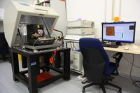
Veeco/Bruker Dimension 3100 AFM
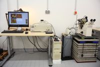
Veeco/Bruker Bioscope AFM
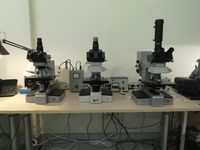
Leitz/Leica Orthoplan microscopes
equipped for brightfield, darkground, phase contrast, polarisation, DIC and fluorescence
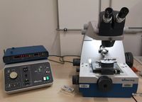
Ultramicrotome Reichert Ultracut
allows ultra-sections down to 50 nm